


Introduction
Kawasaki disease (KD) is a small- to medium-vessel vasculitis diagnosed based on a constellation of clinical features [1]. Atypical presentations of KD are not uncommon, with a reported prevalence of approximately 1.6% in Japan [2]. These atypical presentations may make diagnosis difficult and often delay appropriate treatment. Here, we present an atypical case of KD that manifested with prolonged fever and abdominal pain, initially diagnosed as acalculous cholecystitis.
Case
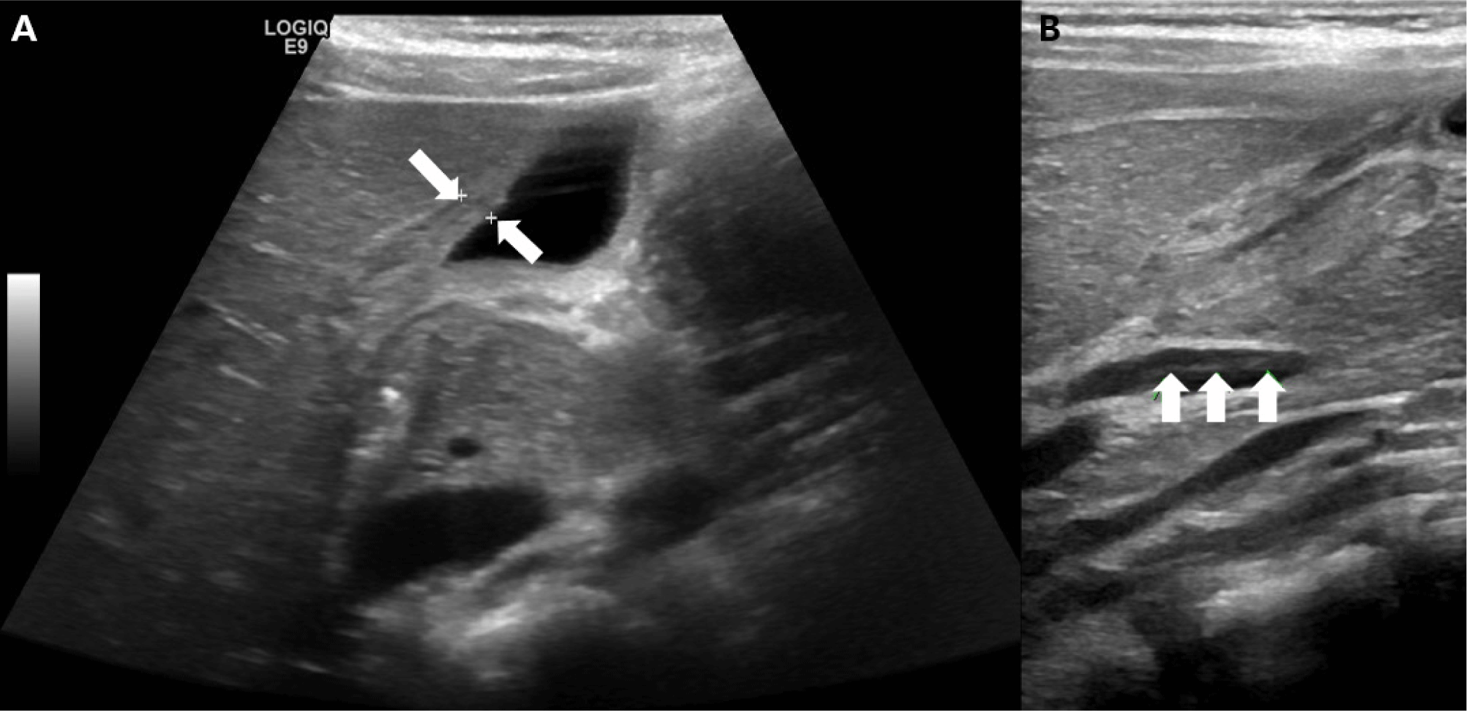
A seven-year-old boy presented to the emergency department with a four-day history of fever and periumbilical abdominal pain that had begun one day earlier. He had vomited once and was awakened by the abdominal pain. The patient had persistent abdominal pain with a severity score of 6/10. The pain did not radiate to the other side or migrate, and it was not related to meals. Physical examination revealed right upper quadrant tenderness, and Murphy’s sign was positive. There were no other clinical features suggestive of KD, except for left cervical lymph node enlargement. Abdominal X-ray revealed non-specific ileus. Laboratory tests showed elevated liver enzymes (total bilirubin, 0.4 mg/dL; aspartate aminotransferase [AST], 269 U/L; alanine aminotransferase [ALT], 134 U/L) and an elevated C-reactive protein (CRP) level of 1.84 mg/dL. The white blood cell (WBC) count was 8,520 /μL (segmented neutrophils 78.0%, lymphocytes 12.6%), hemoglobin 12.7 g/dL, platelet count 238,000 /μL. Respiratory virus detection panel revealed rhino/enterovirus. Based on the patient’s symptoms, physical findings, and laboratory results, enteritis with reactive elevation of liver enzymes due to viral infection or cholecystitis was considered as the differential diagnosis. For differential diagnosis, abdominal ultrasonography was performed. It revealed diffuse wall thickening of the gallbladder (GB) and mild dilatation of the common bile duct with ductal wall thickening (Fig. 1). Inside the common bile duct, a floating echogenic cord-like structure was observed, suggesting biliary sludge (Fig. 1). Ultrasonography revealed no evidence of stones in the GB or bile duct. Based on these findings, acalculous cholecystitis and cholangitis were diagnosed, and the patient was admitted to the gastroenterology department for empirical antibiotic therapy and ursodeoxycholic acid administration.
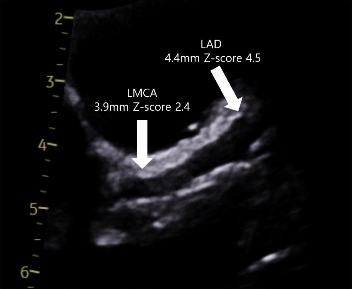
During hospitalization, the patient continued to have a persistent fever despite antibiotic treatment. Except for rhinovirus/enterovirus, all other infection surveillance tests—including blood and stool cultures; viral serologic tests for hepatitis virus, Epstein–Barr virus and herpes simplex virus; diarrhea multiplex polymerase chain reaction (PCR) and the adenovirus antigen test—were negative. On hospital day 3 (the sixth day of fever), he developed bilateral conjunctival injection, a maculopapular rash, and a strawberry tongue. Follow-up laboratory tests showed an interval increase in bilirubin (0.4→4.1 mg/dL) and CRP (1.84→12.94 mg/dL), as well as a marked elevation in NT-proBNP (1,340 pg/mL). The WBC count was 9,660 /μL (segmented neutrophils, 70.1%; lymphocytes, 17.3%), hemoglobin 11.5 g/dL, platelet count 219,000 /μL, albumin 3.3 g/dL, AST/ALT levels 45/285 U/L, and the erythrocyte sedimentation rate 62 mm/hr. Complete KD was diagnosed, and intravenous immunoglobulin (IVIG) and a moderate dose of aspirin (30 mg/kg/day) were initiated. Although multisystem inflammatory syndrome in children could not be fully excluded, the patient tested negative for SARS-CoV-2 by PCR and had no recent history of illness suggestive of SARS-CoV-2 infection. Echocardiography demonstrated borderline dilation of the left main coronary artery (LMCA) (diameter 3.9 mm, Z-score 2.4) and diffuse dilation of the left anterior descending (LAD) coronary artery (diameter 4.4 mm, Z-score 4.5) (Fig. 2). The dimensions of the right coronary artery and left circumflex artery were within the normal range. Z-scores were derived according to the coronary artery Z-score model developed by Yu et al. [3]. In addition, left ventricular contractility was in the lower normal range (ejection fraction 50%–55%), and minimal mitral regurgitation and a minimal amount of pericardial effusion were observed. The treatment was intensified with methylprednisolone at 2 mg/kg/day because of the significantly dilated LAD, in accordance with the updated scientific statement from the American Heart Association [1].
The patient became afebrile 12 hours after the completion of IVIG administration. Although the initial findings suggestive of cholecystitis and cholangitis in this patient might have been related to KD as an extreme manifestation of GB hydrops, we could not exclude the possibility of an incidentally co-occurring disease. For definitive diagnosis and follow-up of these findings, magnetic resonance cholangiopancreatography (MRCP) was performed on hospital day 6. MRCP showed complete resolution of GB wall thickening and bile duct dilatation. On hospital day 8, the patient was discharged on an antiplatelet dose of aspirin (5 mg/kg/day) and a gradual tapering regimen of prednisolone, after echocardiography confirmed no further progression of dilatation in the LMCA and LAD. He was afebrile, and all other features related to KD had completely resolved. His total bilirubin level had normalized, and CRP had decreased from 12.94 to 1.3 mg/dL.
At follow-up in the outpatient clinic, he remained afebrile and had no adverse effects related to the treatment. Desquamation of the extremities was noted. Follow-up echocardiography performed two months after discharge showed a LMCA diameter within the normal range (3.2 mm, Z-score 1.0) and persistent diffuse dilation of the LAD (3.9 mm, Z-score 3.7).
Discussion
There have been few reports of KD presenting as acalculous cholecystitis, and all cases were successfully treated with one or two doses of IVIG [4–8]. The ages of the reported cases ranged from 3 months to 8 years (median 5 years), and all presented with fever accompanied by abdominal pain, vomiting, or diarrhea [4–8]. Two patients also had other features associated with KD at initial presentation [4,8]. Among the five cases described in the literature, three had normal coronary arteries, one showed mild dilation of the LMCA, and one showed mild dilation of both the right and left coronary arteries [6,8]. In this case, the clinical presentation was consistent with previously reported cases, and KD-related features developed during admission. The patient exhibited dilation of LAD.
Gastrointestinal manifestations of KD vary and may include vomiting, diarrhea, cholestatic jaundice, GB hydrops, pancreatitis, and paralytic ileus. Predominant gastrointestinal symptoms have been reported in up to 5% of patients with KD [9]. GB hydrops associated with KD has been reported since the late 1970s, and its self-limiting nature has been well described [10]. In addition, GB hydrops is included as one of the supplementary diagnostic findings for KD in the Japanese guidelines [2]. A few reports have described a higher incidence of coronary complications and intravenous IVIG resistance in patients with sonographic GB abnormalities—such as GB hydrops or acalculous cholecystitis—compared with those without such findings [11,12]. In this case, there was no IVIG resistance, but intensification therapy with corticosteroids was administered. The patient developed significant diffuse dilatation in the LAD, consistent with the previously reported higher incidence of coronary abnormalities in such patients.
The exact mechanism underlying hepatobiliary manifestations in KD remains unclear. Proposed explanations include cystic duct obstruction caused by regional lymphadenopathy or vasculitis of the GB wall, as well as inflammatory infiltration—mechanisms supported by pathological findings from earlier surgically treated cases [12].
In conclusion, this case highlights that KD can initially present as acalculous cholecystitis in the absence of typical clinical features. In patients with prolonged fever and a poor response to conventional cholecystitis therapy, careful evaluation for evolving signs of KD is warranted.