Review

가와사키병에 합병된 대식세포활성증후군: 진단과 치료에 대한 최신 지견

조경순1https://orcid.org/0000-0002-0212-0992, 이수영1,2,*https://orcid.org/0000-0002-5354-3135
Kyoungsoon Cho1https://orcid.org/0000-0002-0212-0992, Soo-Young Lee1,2,*https://orcid.org/0000-0002-5354-3135
Author Information & Copyright
1가톨릭대학교 의과대학 소아과학교실
2가톨릭대학교 의과대학 백신바이오연구소
1Department of Pediatrics, College of Medicine, The Catholic University of Korea, Seoul, Korea
2The Vaccine Bio Research Institute, College of Medicine, The Catholic University of Korea, Seoul, Korea
*Corresponding author: Soo-Young Lee, Department of Pediatrics, Bucheon St. Mary’s Hospital, The Catholic University of Korea, 327 Sosa-ro, Wonmi-gu, Bucheon, Korea, E-mail: sylee@catholic.ac.kr

ⓒ Copyright 2025 Korean Society of Kawasaki Disease. This is an Open-Access article distributed under the terms of the Creative Commons Attribution Non-Commercial License (http://creativecommons.org/licenses/by-nc/4.0/) which permits unrestricted non-commercial use, distribution, and reproduction in any medium, provided the original work is properly cited.

Received: Jan 13, 2025; Revised: Feb 06, 2025; Accepted: Feb 07, 2025

Published Online: Jun 30, 2025

Abstract

Macrophage activation syndrome (MAS), also referred to as secondary hemophagocytic lymphohistiocytosis (HLH), is a hyperinflammatory phenomenon characterized by fever, splenomegaly, cytopenia, and organ dysfunction. In clinical practice, MAS is encountered as a serious complication of systemic inflammatory diseases in children. Although rare, children with Kawasaki disease (KD) may also develop MAS. Compared to KD patients without MAS, those with MAS have more severe laboratory findings and a higher rate of intravenous immunoglobulin resistance and coronary artery abnormalities. The HLH-2004 diagnostic criteria and the 2016 classification criteria are widely used to diagnose KD complicated with MAS (KD-MAS). Hyperferritinemia is the most prominent laboratory finding of KD-MAS, so the ferritin levels should be serially tested in patients suspected of having KD-MAS. Hemophagocytosis is an important histological finding in HLH or MAS, but it can also be seen in critical diseases such as sepsis. For initial treatment of KD-MAS, most institutions use high-dose steroids. Cyclosporine or etoposide is often used to treat steroid-resistant KD-MAS. Recently, cytokine-specific biologics (e.g., anakinra) have been preferred to traditional immunosuppressants in treating pediatric MAS. MAS is easily overlooked in patients with KD due to their similar symptoms. Therefore, it should be considered in KD patients who experience unexpected clinical deterioration despite adequate treatment.

Keywords: Kawasaki Disease; Macrophage Activation Syndrome; Child

서론

가와사키병(Kawasaki disease, KD)은 소아기 급성 전신 혈관염(systemic vasculitis)으로, 발열과 5가지 주요 임상증상(결막염, 구인두의 염증, 발진, 손발의 변화 및 경부 림프절종대)이 함께 나타나는 것이 특징이다[13]. 관상동맥 이상(coronary artery abnormalities, CAAs)은 KD의 가장 중요한 심혈관(cardiovascular) 합병증으로, 치료받지 않는 환자의 15%–25%에서 발생한다[4]. 드물지만 심각한 전신(systemic) 합병증으로, KD 환자에서 대식세포활성증후군(macrophage activation syndrome, MAS)이 발생할 수 있다[5,6].

MAS는 T 세포와 대식세포가 지나치게 활성되어 발생하는 염증 현상으로, 발열, 비장비대, 혈구감소증 및 장기부전(organ dysfunction)을 특징으로 한다[79]. 소아에서 발생하는 MAS의 주요한 원인은 전신형 소아기특발성관절염(systemic juvenile idiopathic arthritis, SJIA)과 전신홍반루프스(systemic lupus erythematosus, SLE)이다[10]. 최근 연구에서 KD에 합병된 MAS (KD complicated with MAS, KD-MAS)에 관한 보고가 증가하면서 KD도 소아 MAS의 주요한 원인에 포함되었다[7]. MAS는 사망률이 30%에 이르는 잠재적으로 치명적인 질환으로, 조기에 진단하여 적극적으로 치료하는 것이 중요하다[8,9]. 하지만 조절되지 않는 발열, 혈소판감소증, 간 기능부전 등 MAS의 주요 임상양상이 심한 형태의 KD에서도 관찰될 수 있으므로, 실제 임상에서 KD의 악화와 MAS의 발생을 구분하기는 쉽지 않다[11,12]. 본 연구에서 저자들은 KD-MAS의 개요에 대해 기술하고 진단과 치료에 도움되는 최신 지견을 제공하려고 한다.

본론

1. 개요
1) 정의와 용어

MAS는 혈구탐식림프조직구증(hemophagocytic lymphohistiocytosis, HLH)의 한 형태이다[13,14]. HLH를 일차성(primary, familial 또는 genetic)과 이차성(secondary, sporadic 또는 acquired)으로 양분하는 분류법은, 복잡한 유전적 인과관계(complex of genetic causality)를 고려하여, 최근에는 지양되는 개념이지만 HLH와 MAS의 정의를 이해하는 데 도움이 된다[15]. 일차성 HLH는 상염색체 열성(autosomal-recessive) 유전 질환으로 대부분 환자들은 영아기에 진단된다[16]. 이차성 HLH는 원인, 즉 유발인자에 따라 감염 연관(infection-associated), 약성종양 연관(malignancy-associated), 류마티스 질환 연관(rheumatic disease-associated), 약물 연관(medication-associated) 등으로 좀 더 세분된다[8,9]. MAS는 1985년 7명의 SJIA 환자에서 처음으로 보고[17]되어 과거에는 류마티스 질환 연관 이차성 HLH로 한정된 용어였으나, 최근 류마티스 질환 외에 다양한 의학적 상황에서도 보고되어 많은 전문가들은 MAS를 이차성 HLH와 동의어로 사용하기도 한다[18].

KD 환자에서 발생된 MAS는 1995년 Ohga et al.[11]이 처음으로 보고하였고, 국내에서는 2002년 Yun et al.[19]이 첫 KD-MAS 사례를 보고하였다. 초기 연구에서는, 다른 원인에 의한 이차성 HLH와 마찬가지로, KD에 합병된 ‘HLH’라고 기술하였으나[20], 최근 연구에서는 대부분 KD에 합병된 ‘MAS’라는 용어를 사용한다[21].

2) 역학과 임상양상

KD 환자에서 MAS의 발생빈도는 북미 연구[22]에서 1.9%(12/638)로, 중국 연구[22]에서는 1.1%(8/719)로 보고하였다. 국내 1개 대학병원의 연구[23]에서 KD-MAS의 발생빈도는 0.8%(4/468)였고[24], 이란은 1.8%(4/218)[25], 인도는 1.3%(12/950)[26]의 발생빈도를 보고하였다. 일본 등 다른 국가의 역학 자료는 아직 보고되지 않았지만, KD(~1.9%)는 이미 SJIA(~10%)와 SLE(~5%) 다음으로 세 번째로 흔한 소아 MAS의 원인으로 간주된다[9,24].

García-Pavón et al.[12]은 체계적 고찰을 통해 69명의 KD-MAS 환자 자료를 분석하였다. 환자의 중앙 연령은 5.6세이었고 남녀 비는 2.1:1(47:22)였다. 대부분 환자(94%)는 KD 진단 후 증상이 악화되면서 나중에 MAS가 진단되었고, 일부 환자(6%)는 MAS가 먼저 진단되고 나중에 KD가 진단되었다. 완전형 KD (complete KD)의 비율은 MAS 동반 없는 KD 환자(75%)[2]와 KD-MAS 환자(77%)가 유사하였다. 이는 KD의 주요 임상증상 유무를 토대로, MAS 동반 없는 KD와 KD-MAS를 구분하기는 어렵다는 것을 의미한다. 하지만, 임상양상의 심한 정도에는 차이를 보인다. 예를 들어 MAS 동반 없는 KD 환자에 비해, KD-MAS 환자에서 정맥주사 면역글로불린(intravenous immunoglobulin, IVIG) 저항성(90%)과 CAAs(46%)가 현저하게 높았다. 또한 빈혈, 중성구감소증, 혈소판감소증, 저알부민혈증, 간효소치 증가, 고페리틴혈증(hyperferritinemia) 등 검사실 소견의 이상도 MAS 동반 없는 KD 환자보다 KD-MAS 환자에서 흔하게 관찰되었다[5].

장기부전은 진단기준에 포함되지 않지만, KD-MAS를 포함한 소아 MAS의 주요 임상양상이다[13]. 소아 MAS의 가장 중요하고 흔한 장기부전은 신경학적 이상과 출혈 성향이며, KD-MAS에서는 심장과 간 기능부전이 추가로 동반될 수 있다[17,20]. 신장 기능부전이 KD-MAS 환자에서 보고되기도 하였다[27].

3) KD와 MAS의 관계

패혈증 환자에서 다양한 형태의 장기부전이 발생하는 것처럼, KD 환자에서 드물지만 심각한 장기부전이 발생할 수 있다[14]. Fig. 1에 나타낸 것처럼, 패혈증 환자에서 확인된 장기부전과 병원체에 따라 패혈성 쇼크(septic shock), 폐렴사슬알균성 중증 패혈증(pneumococcal severe sepsis) 또는 다발기능부전 증후군(multi-organ dysfunction syndrome, MODS)으로 구분할 수 있다[28,29].

kd-3-1-1-g1
Fig. 1. The relationship between MODS and KD-MAS is illustrated. Sepsis plus multi-organ dysfunction is called MODS. Similarly, KD plus can be defined as KD with MODS. MAS is a well-known form of KD with MODS. The figure is modified from Kim et al. [14] with CC-BY-NC. MODS: multi-organ dysfunction syndrome; KD-MAS: Kawasaki disease complicated with macrophage activation syndrome; KD: Kawasaki disease; KDSS: Kawasaki disease shock syndrome; MIS-C: multisystem inflammatory syndrome in children; SARS-CoV-2: severe acute respiratory syndrome coronavirus 2.
Download Original Figure

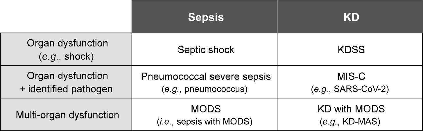
이와 유사하게, KD 환자에서 확인된 장기부전과 병원체에 따라 KD 쇼크증후군(Kawasaki diseases shock syndrome, KDSS), 소아 다기관염증증후군(multisystem inflammatory syndrome in children, MIS-C) 또는 MODS 동반 KD (KD with MODS)로 진단 가능하다[13,14]. 즉, MAS는 KD 환자에서 발생가능한 여러 형태의 MODS 중 하나인 것이다[30]. KDSS, MIS-C 및 KD-MAS의 유사점과 차이점을 이해하는 것은 KD의 주요 임상증상과 사이토카인 폭풍(cytokine storm)을 특징으로 하는 ‘KD 유사 과염증 질환(KD-like hyperinflammatory diseases)’을 연구하는 데 도움이 될 것이다[31,32].

2. 진단
1) 진단기준

KD-MAS을 위한 별도의 진단기준이 없기 때문에, 혈액종양 분과[16]나 류마티스 분과[33]에서 개발한 기준을 차용하여 KD-MAS 환자를 진단한다(Table 1). 2004년 국제 조직구증학회(Histiocyte Society)에서 발표한 HLH-2004 진단기준[16]은 다양한 원인 질환에 합병된 MAS 또는 이차성 HLH의 진단에 광범위하게 이용된다[34,35]. 실제로 지금까지 국내외적으로 발표된 대부분 KD-MAS 연구들은 기본적으로 HLH-2004 기준을 이용하였고, 필요에 따라 다른 진단기준을 추가적으로 활용하였다[6,23]. 그러나 HLH-2004 기준은 원래 혈액종양 질환인 일차성 HLH의 진단을 위해 개발된 것으로, SJIA나 KD 환자에서 발생한 이차성 HLH를 진단하기에는 민감도(sensitivity)가 낮다[25,26]. 이러한 제한점을 보완하기 위해, 2016년 소아류마티스 국제 임상시험기구(Paediatric Rheumatology International Trials Organisation, PRINTO)에서 SJIA 환자를 위한 2016 MAS 분류기준[33]을 제안하였다. 2016 MAS 기준은 SJIA뿐만 아니라, SLE, KD 등 다양한 자가면역(autoimmune) 질환과 자가염증(autoinflammatory) 질환에 합병된 MAS 진단에 유용하다고 알려졌다[36].

Table 1. KD-MAS diagnosis: HLH-2004 diagnostic criteria [16] and 2016 MAS classification criteria [33]
HLH-2004 2016 MAS classification
Fever -
Splenomegaly -
Cytopenia ≥ 2 cell lines -
 Hemoglobin < 9.0 g/dL -
 Neutrophils < 1,000 /μL (Any two of the following four criteria)
 Platelets < 100,000 /μL  i) Platelet ≤ 181,000 /μL
(Elevated liver enzymes are supportive evidence)  ii) AST > 48 U/L
Triglycerides ≥ 265 mg/dL  iii) Triglycerides > 156 mg/dL
 or fibrinogen ≤ 150 mg/dL  iv) Fibrinogen ≤ 360 mg/dL
Ferritin ≥ 500 ng/mL Ferritin > 684 ng/mL (essential)
sCD25 (soluble IL-2 receptors) ≥ 2,400 U/mL -
Decrease or absent NK cell activity -
Hemophagocytosis in bone marrow, liver, or lymph nodes -
Diagnosed when ≥ 5/8 criteria are met Diagnosed when hyperferritinemia + ≥ 2/4 criteria

KD-MAS: Kawasaki disease complicated with macrophage activation syndrome; HLH: hemophagocytic lymphohistiocytosis; MAS: macrophage activation syndrome; -: not applicable; AST: aspartate aminotransferase; IL-2: interleukin-2; NK: natural killer.

Download Excel Table

그 밖에 혈구탐식증후군 진단점수(hemophagocytic syndrome diagnostic score, HScore)[37], SJIA 환자의 MAS 점수(MAS in patients with SJIA score; MS score)[38] 등을 KD-MAS의 진단에 적용할 수 있다. 각 진단 방법은 서로 경쟁적이라기 보다는 보완적이기 때문에, 임상 상황에 따라 적절한 방법을 선택한다[10]. 일부 연구자들은 진단 정확성을 높이기도 위해 복수의 진단 방법을 이용하기도 한다[13,23].

2) 진단적 단서 또는 함정
(1) 고페리틴혈증

페리틴 수치가 급증하는 것은 MAS의 가장 중요한 검사실 소견이다[21,39]. 따라서, 고페리틴혈증은 모든 MAS 진단기준에 포함되었다. 페리틴 수치 500 ng/mL 이상이 최소 기준이지만, KD-MAS 환자의 페리틴 수치는 대부분 1,000 ng/mL 이상이며 10,000 ng/mL 이상인 경우도 드물지 않다[40]. Eloseily et al.[41]은 페리틴/erythrocyte sedimentation rate (ESR) 비율의 증가(ferritin/ESR ratio ≥ 21.5)가 MAS의 조기 진단에 유용하다(민감도 82%, 특이도 78%)고 제안하였다. 페리틴 수치는 질병의 활성도, 치료 반응 및 예후의 예측에도 활용된다[34,35].

(2) 혈구탐식구증(hemophagocytosis)

골수검사에서 혈구탐식구증을 확인하는 것은 HLH 또는 MAS의 진단을 강력하게 지지해주는 조직학적 소견이다[42]. 하지만, 혈구탐식구증은 HLH나 MAS의 질병특유(pathognomonic) 병변이 아니며, 패혈증과 같은 심한 전신염증을 유발하는 질환에서도 관찰된다[43]. 반면에 혈구탐식구증이 모든 MAS 환자에서 입증되지도 않는다. 인도의 소아 MAS 연구[44]에서 절반 이상(18/31)의 환자들은 혈구탐식구증 확인 없이 혈구감소증, 고페리틴혈증 등 다른 기준을 5가지 이상 만족하여 MAS로 진단되었다. 즉, 혈구탐식구증은 MAS 진단의 필요 조건이나 충분 조건은 아닌 것이다[13]. 따라서, Gupta et al.[45]은 혈구탐식구증을 확인하지 못했다는 이유로 MAS의 진단이나 치료를 지연해서는 안 된다고 강조하였다.

(3) CAAs

심장초음파 검사에서 전형적인 CAAs를 확인하는 것이 KD-MAS 진단 지연의 원인이 되기도 한다. 이는 CAAs를 KD의 특이적 소견으로 간주하여 KD 외에 다른 질환을 임상적으로 의심하지 못하였기 때문이다[10]. 하지만, CAAs는 심근염, 타카야수 혈관염(Takayasu’s arteritis), SJIA, 류마티스 열(rheumatic fever), SLE 등, 심한 염증반응을 유발하는 다양한 소아기 질환에서 관찰된다[46]. 즉 CAAs는 KD의 진단을 지지해 주는 소견이기도 하지만, 심한 염증반응의 지표일 수도 있다[47]. 따라서 혈구탐식구증이나 CAAs의 유무와 무관하게, KD 환자가 적절한 치료에도 불구하고 예상치 못한 심한 임상양상을 나타낼 때 MAS에 대한 선별검사(혈소판, 간효소, 페리틴, 중성지방, 피브리노겐 등)를 시행해야 한다.

3) 잠재적 MAS

다양한 전신염증 질환에서 MAS의 발생을 조기 인식하기 위해, 잠재적 MAS의 개념이 유용하다. ‘잠재적(occult 또는 subclinical) MAS’는 현 시점에서 MAS 진단기준을 만족하지 못하지만, 임상양상이 악화되면 ‘명시적(overt 또는 fulminant) MAS’로 진행가능한 염증상태를 의미한다[48,49]. 잠재적 MAS 환자에서 MAS를 완전히 배제하지 말고 MAS의 발생 가능성을 임상적으로 의심하는 것이 중요하다[49]. 이는 당뇨 전 단계(pre-diabetic) 환자를 면밀하게 추적 관찰하여 당뇨로 진행되는 것을 조기에 진단하고 치료하는 것과 유사한 원리이다.

Jeong et al.[5]은 KD의 일반적이지 않은 임상 상황, 즉 IVIG 저항성, 비장비대, 혈소판감소증, 고페리틴혈증 또는 장기부전을 나타내는 환자를 MAS 발생 고위험군(즉, 잠재적 MAS)으로 간주하여 세심하게 관찰해야 한다고 강조하였다. 실제로, 고위험군으로 간주되는 IVIG 저항성 KD 환자의 MAS 발생빈도(~12%)는 일반 KD 환자의 MAS 발생빈도(~1.9%)보다 6배 이상으로 높다[24].

3. 치료
1) 기본 원칙

지금까지 소아 MAS 치료지침의 개발을 위해 시행된 임상 연구가 없기 때문에, KD-MAS를 포함한 대부분의 소아 MAS 환자는 일차성 HLH 치료지침과 이전의 MAS 치료경험을 토대로 치료를 받는다[8]. 소아 MAS 치료의 기본 원칙은 i) 원인 질환 또는 유발인자의 관리, ii) 수액, 전해질, 영양 보충, 수혈 등 지지요법(supportive care) 및 iii) 조절에서 벗어난 염증반응을 완화시키는 것이다[50]. 국내외 연구[5,12]에서, 모든 KD-MAS 환자들은 기본적으로 IVIG와 아스피린(즉, KD 표준치료) 치료를 받았다. MAS 동반 없는 KD 환자와 동일하게, CAAs의 정도에 따라 다른 종류의 항혈소판제나 항응고제를 추가할 수 있다[1,50]. 또한, 심한 출혈성향을 보이는 KD-MAS 환자에서는 프로트롬빈 시간의 국제 표준화 비율(international normalized ratio of prothrombin time, INR)을 참고하여 항혈전요법을 조절해야 한다[50].

대부분의 KD-MAS 환자들은 KD 표준치료에도 불구하고 발열이 지속되어 추가적인 치료가 필요하다[21,24]. 첫 번째 IVIG에 저항성을 보이는 KD-MAS 치료를 위해 가장 많이 사용하는 약제는 고용량 정맥주사 스테로이드(intravenous methylprednisolone [IVMP] 30 mg/kg/day for 3–5 days)이다[5,12]. IVMP와 두 번째 IVIG를 함께 치료하기도 한다[51]. 스테로이드 저항성 KD-MAS의 치료방법은 기관에 따라 다르다.

2) 치료약제: 면역조절제 또는 생물학적 제제

많은 기관에서 스테로이드 저항성 KD-MAS 치료를 위해 사이클로스포린(cyclosporine)을 사용한다. 국내(74%, 17/23)[5]와 국외(49%, 34/69)[12] 보고에 따르면, 사이클로스포린은 KD-MAS의 치료에서 스테로이드 다음으로 많이 사용된 약제였다. 일부 전문가는 KD-MAS의 일차 치료에 IVMP와 사이클로스포린을 병합하여 사용한다[9]. 혈액종양 분과에서는 사이클로스포린보다 에토포시드(etoposide)를 선호한다[16]. 최근 미국의 의료기관에서는 소아 MAS의 일차 또는 이차 치료를 위해 사이클로스포린이나 에토포시드와 같은 비특이적 면역조절제보다 사이토카인 특이적(cytokine-specific) 생물학적 제제(anakinra 또는 tocilizumab)를 많이 사용한다[52,53]. 국외 KD-MAS 환자에 비해, 국내 KD-MAS 환자에서 사이클로스포린과 에토포시드의 치료 비율이 높았고 생물학적 제제의 치료 비율은 낮았다(Table 2).

Table 2. KD-MAS treatment: systematic review [12] and Korean study [5]
Variables Systematic review [n = 69 (%)] Korean study [n = 23 (%)]
IVIG and aspirin 69 (100) 23 (100)
Systemic steroids 60 (87) 23 (100)
Cyclosporine 34 (49) 17 (74)
Etoposide 27 (39) 16 (70)
Biologics (anti-TNF, IL-1, or IL-6) 7 (10) 1 (4)
HLH-2004 protocol1) 24 (35) 15 (65)

1) The HLH-2004 protocol consists of 40 weeks of combination chemotherapy including etoposide and cyclosporine.

KD-MAS: Kawasaki disease complicated with macrophage activation syndrome; IVIG: intravenous immunoglobulin; TNF: tumor necrosis factor; IL: interleukin; HLH: hemophagocytic lymphohistiocytosis.

Download Excel Table
3) 치료기간: 8주 또는 40주

KD-MAS 환자의 치료반응을 적절하게 추적 관찰하는 것이 중요하다. 질병 활성도 평가를 위해, 페리틴, 응고인자, 백혈구, 혈소판, 간효소 등에 대한 검사를 반복적으로 시행해야 한다[15,34]. 압도적인 임상양상으로 인해, KD-MAS 환자들은 종종 필요 이상의 치료(over-treatment)를 받기도 한다[54]. 국내외 KD-MAS 치료 현황의 비교에서 가장 큰 차이점은 국내 환자들이 높은 비율(65%)로 HLH-2004 치료지침, 즉 에토포시드를 포함하는 40주 이상의 복합 항암치료(combination chemotherapy)를 받았다는 것이다(Table 2). 이는 국내 KD-MAS 환자에서 필요 이상의 치료가 시행되었을 가능성을 암시한다.

Haytoglu et al.[55]은 소아 MAS 환자의 약 80%는 8주 동안의 단순 면역조절제 치료로 완전 관해(complete remission)되었다고 보고했다. MAS 진단시의 심한 양상양상이 40주 이상의 복합 항암치료가 필요하다는 것을 의미하지는 않는다. 따라서, MAS 치료 초기 2-6주 동안의 질병 활성도를 적절하게 평가하여 최종적인 치료 기간과 약제를 신중하게 결정해야 한다[15,34]. 향후 국내외 공동 연구를 통해, KD-MAS 환자에게 보편적으로 적용할 수 있는 치료지침의 개발이 필요할 것이다.

결론

MAS는 KD 환자에서 발생할 수 있는 여러 형태의 장기부전 중 하나이다. KD-MAS를 간과하지 않기 위해서는 임상적으로 의심하는 것이 중요하다. 예를 들어 IVIG 저항성, 비장비대, 혈소판감소증, 고페리틴혈증, 장기부전과 같이, KD에서 일반적이지 않은 임상 상황에서 MAS의 발생 가능성을 염두에 두어야 한다. KD-MAS의 진단에는 HLH-2004 기준, 2016 MAS 기준 등을 사용한다. 각 진단 방법은 장단점이 있으므로, 상황에 따라 적절한 기준을 선택한다. KD-MAS의 일차 치료에는 고용량 IVMP를 사용하며, 스테로이드 저항성 KD-MAS에는 사이클로스포린, 에토포시드 및 생물학적 제제를 적용할 수 있다. 최근에는 소아 MAS의 치료에는 전통적인 면역억제제보다 생물학적 제제를 선호하는 추세이다. 아직까지 KD-MAS를 포함한 소아 MAS의 진단과 치료를 위해 발표된 진료지침이 없다. 추후 연구를 통해 KD-MAS 환자에게 보편적으로 적용할 수 있는 진료지침의 개발이 요구된다.

Conflict of Interest

No potential conflict of interest relevant to this article was reported.

Funding

This study was supported by the Institute of Clinical Medicine Research of Bucheon St. Mary’s Hospital, Research Fund, 2025.

Acknowledgements

Not applicable.

Authors’ Contributions

Conceptualization: Lee SY.

Data curation: Cho K.

Formal analysis: Lee SY.

Methodology: Cho K.

Software: Cho K, Lee SY.

Validation: Cho K.

Investigation: Lee SY.

Writing - original draft: Cho K.

Writing - review & editing: Cho K, Lee SY.

Ethics Approval

Not applicable.

References

1.

McCrindle BW, Rowley AH, Newburger JW, Burns JC, Bolger AF, Gewitz M, et al. Diagnosis, treatment, and long-term management of Kawasaki disease: a scientific statement for health professionals from the American Heart Association. Circulation. 2017; 135:e927-99

2.

Han SB, Lee SY. Differentiating Kawasaki disease from urinary tract infection in febrile children with pyuria and C-reactive protein elevation. Ital J Pediatr. 2018; 44-137

3.

Kim GB, Park S, Eun LY, Han JW, Lee SY, Yoon KL, et al. Epidemiology and clinical features of Kawasaki disease in South Korea, 2012–2014. Pediatr Infect Dis J. 2017; 36:482-5

4.

Newburger JW, Takahashi M, Burns JC, Beiser AS, Chung KJ, Duffy CE, et al. The treatment of Kawasaki syndrome with intravenous gamma globulin. N Engl J Med. 1986; 315:341-7

5.

Jeong DC, Lee SY. Macrophage activation syndrome in Kawasaki disease: a literature review of Korean studies. J Rheum Dis. 2025; 32:105-12

6.

Natoli V, Rosina S, Ravelli A. Is macrophage activation syndrome in Kawasaki disease underrecognized?. J Rheumatol. 2021; 48:162-4

7.

Cron RQ, Davi S, Minoia F, Ravelli A. Clinical features and correct diagnosis of macrophage activation syndrome. Expert Rev Clin Immunol. 2015; 11:1043-53

8.

Ravelli A, Davì S, Minoia F, Martini A, Cron RQ. Macrophage activation syndrome. Hematol Oncol Clin North Am. 2015; 29:927-41

9.

Crayne C, Cron RQ. Pediatric macrophage activation syndrome, recognizing the tip of the Iceberg. Eur J Rheumatol. 2020; 7:S13-20

10.

Lee J, Bae KS, Rhim JW, Lee SY, Jeong DC, Kang JH. Macrophage activation syndrome in children: update on diagnosis and treatment. Children. 2024; 11-755

11.

Ohga S, Ooshima A, Fukushige J, Ueda K. Histiocytic haemophagocytosis in a patient with Kawasaki disease: changes in the hypercytokinaemic state. Eur J Pediatr. 1995; 154:539-41

12.

García-Pavón S, Yamazaki-Nakashimada MA, Báez M, Borjas-Aguilar KL, Murata C. Kawasaki disease complicated with macrophage activation syndrome: a systematic review. J Pediatr Hematol Oncol. 2017; 39:445-51

13.

Han SB, Lee SY. Macrophage activation syndrome in children with Kawasaki disease: diagnostic and therapeutic approaches. World J Pediatr. 2020; 16:566-74

14.

Kim BJ, Kim D, Cho K, Rhim JW, Lee SY, Jeong DC. Kawasaki disease shock syndrome and macrophage activation syndrome: a case report. Kawasaki Dis. 2023; 1e11

15.

Jordan MB, Allen CE, Greenberg J, Henry M, Hermiston ML, Kumar A, et al. Challenges in the diagnosis of hemophagocytic lymphohistiocytosis: recommendations from the North American Consortium for Histiocytosis (NACHO). Pediatr Blood Cancer. 2019; 66e27929

16.

Henter JI, Horne A, Aricó M, Egeler RM, Filipovich AH, Imashuku S, et al. HLH-2004: diagnostic and therapeutic guidelines for hemophagocytic lymphohistiocytosis. Pediatr Blood Cancer. 2007; 48:124-31

17.

Hadchouel M, Prieur AM, Griscelli C. Acute hemorrhagic, hepatic, and neurologic manifestations in juvenile rheumatoid arthritis: possible relationship to drugs or infection. J Pediatr. 1985; 41:561-6

18.

Grom AA. Macrophage activation syndrome and reactive hemophagocytic lymphohistiocytosis: the same entities?. Curr Opin Rheumatol. 2003; 15:587-90

19.

Yun HJ, Jeon KW, Kim HM, Park SW, Uh Y. Hemophagocytic syndrome with Kawasaki disease and peripheral gangrene. J Korean Pediatr Soc. 2002; 45:664-8.

20.

Kim HK, Kim HG, Cho SJ, Hong YM, Sohn S, Yoo ES, et al. Clinical characteristics of hemophagocytic lymphohistiocytosis related to Kawasaki disease. Pediatr Hematol Oncol. 2011; 28:230-6

21.

Roh DE, Kwon JE, Choi HJ, Kim YH. Importance of serum ferritin level for early diagnosis and differentiation in patients with Kawasaki disease with macrophage activation syndrome. Children. 2021; 8-269

22.

Latino GA, Manlhiot C, Yeung RSM, Chahal N, McCrindle BW. Macrophage activation syndrome in the acute phase of Kawasaki disease. J Pediatr Hematol Oncol. 2010; 32:527-31

23.

Wang W, Gong F, Zhu W, Fu S, Zhang Q. Macrophage activation syndrome in Kawasaki disease: more common than we thought?. Semin Arthritis Rheum. 2015; 44:405-10

24.

Rhee S, Kim D, Cho K, Rhim JW, Lee SY, Jeong DC. Under-recognized macrophage activation syndrome in refractory Kawasaki disease: a wolf in sheep’s clothing. Children. 2022; 9-1588

25.

Mousavi MS, Assari R, Tahghighi F, Eshaghi H, Ziaee V. Prolonged fever and intravenous immunoglobulin resistance in Kawasaki disease: should macrophage activation syndrome be considered?. Iran J Pediatr. 2019; 29e69170

26.

Pilania RK, Jindal AK, Johnson N, Prithvi A, Vignesh P, Suri D, et al. Macrophage activation syndrome in children with Kawasaki disease: an experience from a tertiary care hospital in northwest India. Rheumatology. 2021; 60:3413-9

27.

Chuang GT, Tsai IJ, Lin MT, Chang LY. Acute kidney injury in patients with Kawasaki disease. Pediatr Res. 2016; 80:224-7

28.

Goldstein B, Giroir B, Randolph A. International pediatric sepsis consensus conference: definitions for sepsis and organ dysfunction in pediatrics. Pediatr Crit Care Med. 2005; 6:2-8

29.

Lee S, Kim D, Kim BJ, Rhim JW, Lee SY, Jeong DC. Comparison of COVID-19-associated multisystem inflammatory syndrome in children (MIS-C) and Kawasaki disease shock syndrome: case reports and literature review. J Rheum Dis. 2023; 30:272-7

30.

Han SB, Lee SY. The link between Kawasaki disease shock syndrome and macrophage activation syndrome in terms of organ dysfunction observed in children with systemic inflammatory response syndrome. Paediatr Int Child Health. 2018; 38:308-9

31.

Lee J, Kim BJ, Cho KS, Rhim JW, Lee SY, Jeong DC. Similarities and differences between multisystem inflammatory syndrome in children (MIS-C) and Kawasaki disease shock syndrome. Children. 2023; 10-1527

32.

Lee J, Lee SY. Under-recognised macrophage activation syndrome in multisystem inflammatory syndrome in children. Clin Exp Rheumatol. 2024; 42:2096-7

33.

Ravelli A, Minoia F, Davì S, Horne AC, Bovis F, Pistorio A, et al. 2016 Classification criteria for macrophage activation syndrome complicating systemic juvenile idiopathic arthritis: a European league against rheumatism/American College of Rheumatology/Paediatric Rheumatology International Trials Organisation Collaborative Initiative. Ann Rheum Dis. 2016; 75:481-9

34.

Griffin G, Shenoi S, Hughes GC. Hemophagocytic lymphohistiocytosis: an update on pathogenesis, diagnosis, and therapy. Best Pract Res Clin Rheumatol. 2020; 34-101515

35.

Sztajnbok F, Fonseca AR, Campos LR, Lino K, Rodrigues MCF, Silva RM, et al. Hemophagocytic lymphohistiocytosis and macrophage activation syndrome: two rare sides of the same devastating coin. Adv Rheumatol. 2024; 64-28

36.

Han SB, Lee SY, Jeong DC, Kang JH. Should 2016 criteria for macrophage activation syndrome be applied in children with Kawasaki disease, as well as with systemic-onset juvenile idiopathic arthritis?. Ann Rheum Dis. 2016; 75:E44

37.

Fardet L, Galicier L, Lambotte O, Marzac C, Aumont C, Chahwan D, et al. Development and validation of the HScore, a score for the diagnosis of reactive hemophagocytic syndrome. Arthritis Rheumatol. 2014; 66:2613-20

38.

Minoia F, Bovis F, Davì S, Horne AC, Fischbach M, Frosch M, et al. Development and initial validation of the MS score for diagnosis of macrophage activation syndrome in systemic juvenile idiopathic arthritis. Ann Rheum Dis. 2019; 78:1357-62

39.

Chen Y, Shang S, Zhang C, Liu T, Yang Z, Tang Y. Hemophagocytic lymphohistiocytosis at initiation of Kawasaki disease and their differential diagnosis. Pediatr Hematol Oncol. 2010; 27:244-9

40.

Allen CE, Yu X, Kozinetz CA, McClain KL. Highly elevated ferritin levels and the diagnosis of hemophagocytic lymphohistiocytosis. Pediatr Blood Cancer. 2008; 50:1227-35

41.

Eloseily EMA, Minoia F, Crayne CB, Beukelman T, Ravelli A, Cron RQ. Ferritin to erythrocyte sedimentation rate ratio: simple measure to identify macrophage activation syndrome in systemic juvenile idiopathic arthritis. ACR Open Rheumatol. 2019; 1:345-9

42.

Nair V, Das S, Sharma A, Sharma S, Sharma P, Ray S, et al. A clinicopathological analysis of 26 patients with infection-associated haemophagocytic lymphohistiocytosis and the importance of bone marrow phagocytosis for the early initiation of immunomodulatory treatment. Postgrad Med J. 2013; 89:185-92

43.

Castillo L, Carcillo J. Secondary hemophagocytic lymphohistiocytosis and severe sepsis/systemic inflammatory response syndrome/multiorgan dysfunction syndrome/macrophage activation syndrome share common intermediate phenotypes on a spectrum of inflammation. Pediatr Crit Care Med. 2009; 10:387-92

44.

Pal P, Bathia J, Giri PP, Roy M, Nandi A. Macrophage activation syndrome in pediatrics: 10 years data from an Indian center. Int J Rheum Dis. 2020; 23:1412-6

45.

Gupta A, Weitzman S, Abdelhaleem M. The role of hemophagocytosis in bone marrow aspirates in the diagnosis of hemophagocytic lymphohistiocytosis. Pediatr Blood Cancer. 2008; 50:192-4

46.

Kim JH, Kim HJ, Shin JH, Choi UY, Lee SY, Han JW. Coronary arterial lesions of Kawasaki disease observed in a mouse model of sepsis: a pilot study and a review of the literature. Pediatr Infect Vaccine. 2017; 24:102-7

47.

Han SB, Lee SY, Jeong DC. The presence of coronary artery lesions may indicate the severity of the inflammation rather than the cause. Semin Arthritis Rheum. 2017; 47e6

48.

Behrens EM, Beukelman T, Paessler M, Cron RQ. Occult macrophage activation syndrome in patients with systemic juvenile idiopathic arthritis. J Rheumatol. 2007; 34:1133-8.

49.

Choi UY, Han SB, Lee SY, Jeong DC. Should refractory Kawasaki disease be considered occult macrophage activation syndrome?. Semin Arthritis Rheum. 2017; 46e17

50.

Yoo BW. Antithrombotic therapy in Kawasaki disease. Kawasaki Dis. 2023; 1e15

51.

Bagri NK, Gupta L, Sen ES, Ramanan AV. Macrophage activation syndrome in children: diagnosis and management. Indian Pediatr. 2021; 58:1155-61

52.

Grom AA, Horne AC, De Benedetti F. Macrophage activation syndrome in the era of biologic therapy. Nat Rev Rheumatol. 2016; 12:259-68

53.

Eloseily EM, Weiser P, Crayne CB, Haines H, Mannion ML, Stoll ML, et al. Benefit of anakinra in treating pediatric secondary hemophagocytic lymphohistiocytosis. Arthritis Rheumatol. 2020; 72:326-34

54.

Han SB, Lee SY. Children with extreme hyperferritinemia are at risk of receiving more chemotherapy than necessary. Turk J Pediatr. 2020; 62:165-6

55.

Haytoglu Z, Yazici N, Erbay A. Secondary hemophagocytic lymphohistiocytosis: do we really need chemotherapeutics for all patients?. J Pediatr Hematol Oncol. 2017; 39:e106-9