

Introduction
Kawasaki disease (KD), first described in 1967, is an acute systemic vasculitis predominantly affecting infants and young children. Although most patients recover without sequelae, coronary artery involvement remains the most clinically significant complication. With the widespread adoption of high-dose intravenous immunoglobulin, the incidence of coronary aneurysms has decreased substantially; however, giant aneurysms and progressive coronary stenosis persist in a small but high-risk subgroup [1].
KD-related coronary sequelae include aneurysm formation, intimal proliferation, calcification, thrombosis, myocardial infarction (MI), and are often accompanied by robust collateral formation. These lesions frequently involve the proximal coronary arteries, especially the left main trunk and left anterior descending (LAD) artery, and complicate ischemia detection and decision-making regarding revascularization [2]. As children survive into adolescence and adulthood, the long-term burden of coronary pathology necessitates durable revascularization strategies. Prior to the 1970s, pediatric coronary artery bypass surgery was rarely performed. The emergence of KD-related coronary sequelae prompted development of pediatric revascularization techniques, ultimately establishing coronary artery bypass grafting (CABG) as a definitive therapeutic option [3]. This review summarizes the evolution of CABG in KD-related coronary sequelae, with emphasis on conduit selection, graft biology, long-term patency, survival, and contemporary surgical principles.
Main Subject
KD induces a monophasic vasculitis affecting medium-sized muscular arteries, especially the coronary arteries. During the acute phase, pan-vasculitis disrupts elastic lamina integrity, leading to aneurysm formation [1]. Giant aneurysms (≥ 8 mm) are particularly prone to thrombosis and early MI. In the convalescent phase, intimal proliferation and fibrocellular remodeling lead to progressive stenosis, typically at aneurysm inlet or outlet segments [4]. Calcification is common in late adolescence and adulthood.
Selective coronary angiography in KD revealed that obstructive lesions typically occur at the proximal inlet or outlet of aneurysmal segments. Lesions are concentrated in the left main trunk and proximal LAD, although right coronary artery (RCA) involvement is also common. Children frequently develop extensive intercoronary and intracoronary collateral vessels. As a result, ischemic symptoms may be less prominent than in adults despite severe anatomical stenosis [3]. However, MI remains a major determinant of morbidity and mortality. Historical reports demonstrated markedly increased mortality after repeated infarctions, highlighting the need for definitive revascularization in selected patients [5,6]. Distinct features of KD-related coronary sequelae include predominant proximal LAD and left main involvement, multivessel disease in giant aneurysm cases, calcified and aneurysm-associated stenosis, well-developed collateral circulation, and variable regression or progression over time [4]. These characteristics distinguish KD-related coronary sequelae from adult atherosclerosis and necessitate disease-specific management strategies.
Surgical indications gradually became accepted as experience accumulated, particularly in patients with severe proximal stenosis, multivessel involvement, recurrent thrombosis, impaired left ventricular function, and previous MI. Importantly, many children remain asymptomatic despite severe stenosis due to collateral circulation. Therefore, objective ischemia documentation is essential [3].
The first pediatric CABG procedures for KD-related coronary sequelae were performed using autologous saphenous vein grafts (SVGs) [7]. Early postoperative angiography demonstrated satisfactory patency and improvement in left ventricular ejection fraction (LVEF). However, long-term outcomes were disappointing. SVGs frequently occluded within several years, often due to intimal hyperplasia, thrombosis, and progressive degeneration. In addition, vein grafts lacked growth potential, resulting in mismatch between graft length and somatic growth of the child. This occasionally led to traction, kinking, and compromised distal runoff. Long-term follow-up demonstrated significantly inferior patency of SVGs compared with arterial grafts. Twenty-year patency rate was approximately 44% for SVGs, while it was 87% for internal thoracic artery (ITA) grafts. The disparity was even more pronounced in children younger than 10 years at the time of operation [8]. These findings established that SVGs are suboptimal conduits in growing children and should generally be avoided.
The introduction of the ITA graft as a conduit in pediatric CABG represented a transformative advance. The ITA graft possesses structural characteristics distinct from muscular arteries. It contains more elastic fibers and fewer smooth muscle cells, rendering it relatively resistant to inflammatory changes associated with KD, and also has reduced atherosclerotic degeneration. In 1983, ITA grafting was successfully performed in children with KD-related coronary sequelae [9]. Subsequent experience demonstrated excellent patency, structural stability, and compatibility with growth. Bilateral ITA grafting was shown to be safe and did not impair thoracic development. The ITA-based approach became the global standard and is often referred to as the “Kitamura operation.” [3,8,10–13].
One of the most important observations in pediatric CABG is the growth capacity of ITA grafts. Serial angiographic analyses demonstrated significant longitudinal and circumferential growth proportional to increases in body surface area. Mathematical modeling revealed a strong correlation between ITA graft length and somatic growth (correlation coefficient r = 0.845, P = 0.001). Circumferential growth was also documented, with increasing graft diameter over time without degenerative wall changes [14,15]. In contrast, SVGs showed no significant correlation with body surface area and exhibited degenerative changes including wall irregularity, dilation, and fibroproliferative thickening. The durability of ITA grafts to grow harmoniously with the child ensures sustained hemodynamic compatibility over decades, a critical consideration given the long-life expectancy of these patients.
Longitudinal studies extending up to 20–30 years provide robust evidence of ITA superiority. The 20-year ITA graft patency rate is approximately 87%, whereas the SVG shows 44% patency rate at the same period. The ITA grafts also show significantly superior patency rate for both LAD and non-LAD target vessels, and no significant difference in the patency rate between children younger and older than 10 years. Furthermore, ITA grafts that were patent at one year postoperatively generally remained patent over long-term follow-up. In contrast, SVG failure occurred both early and late, frequently associated with intimal hyperplasia and atherosclerotic degeneration. These findings strongly support exclusive or predominant use of arterial grafts in pediatric revascularization [8].
The ITA string phenomenon may occur when competitive native coronary flow reduces graft flow. This can result in narrowing or functional occlusion of the graft. Interestingly, spontaneous recanalization of ITA grafts has been observed in approximately 20%–25% of cases, particularly when progression of native stenosis reduces competitive flow [16]. Fractional flow reserve measurement has been proposed as a tool to guide surgical decision-making and potentially reduce competitive flow-related graft failure [17,18]. However, its role in KD requires further prospective evaluation.
Long-term survival following ITA-based CABG for KD-related coronary sequelae is excellent. Thirty-year survival rates approach 95%. Surgical mortality has been reported as negligible in experienced centers. Approximately one-third of patients had prior MI at the time of surgery; nevertheless, survival remained favorable when ventricular function was preserved. However, cardiac event-free survival declines gradually over time, with reported rates of approximately 60%–70% at 25 years. Cardiac events include graft stenosis, progression of native coronary disease, new obstructive lesions, acute MI, arrhythmias, requirement for further intervention (percutaneous coronary intervention [PCI] or reoperation), and deaths. Late mortality is often associated with reduced LVEF and ventricular tachyarrhythmias rather than graft failure. These findings emphasize the importance of early intervention before irreversible myocardial damage occurs [3,8].
Pediatric ITAs are small-caliber vessels, sometimes measuring less than 1 mm in diameter in infants. Meticulous microsurgical technique is therefore essential. The key technical elements include use of surgical microscopes or high-magnification loupes, fine monofilament sutures (8-0 or 9-0 sutures), precise anastomotic technique, and usually pedicled ITA harvesting. The use of microsurgical methods has expended indications to include infants and very small children [3].
Anastomosis stenosis may occur but can often be managed successfully with balloon angioplasty [19]. Coronary stenting is generally avoided in children due to vessel size and long-term considerations.
PCI represents an alternative revascularization strategy in selected patients with KD-related coronary sequelae, although its role remains limited compared with CABG. Unlike surgical bypass, PCI targets the native coronary artery and is therefore more susceptible to restenosis and the need for repeat intervention [1]. The unique pathological features of KD-related coronary sequelae―including severe calcification, aneurysmal segments, and luminal myofibroblastic proliferation―pose significant technical challenges [20].
Available PCI modalities include balloon angioplasty, stent implantation, and adjunctive techniques such as rotational atherectomy and intravascular lithotripsy; however, procedural risks such as neo-aneurysm formation and restenosis remain concerns, with reported restenosis rates of approximately 20%–25% following angioplasty [20,21]. Clinical data suggest that PCI may achieve favorable short-term outcomes in localized lesions, especially in older children or adults, but is associated with higher rates of reintervention compared with CABG [22]. Consequently, PCI is generally reserved for carefully selected cases, such as focal stenosis or acute MI requiring urgent reperfusion, whereas CABG remains the preferred strategy for multivessel disease and complex coronary anatomy in KD-related coronary sequelae, particularly in small children.
Contemporary surgical strategies emphasize exclusive or predominant arterial revascularization, bilateral ITA grafting when anatomically feasible, avoidance of SVGs in children, complete revascularization of significant lesions [23], and long-term multidisciplinary follow-up. As patients age into adulthood, reintervention involve radial artery grafting, gastroepiploic artery use [24], or PCI in larger vessels [22]. Cardiac transplantation remains a rare but necessary option in cases of severe ventricular dysfunction and diffuse coronary pathology.
The evolution of CABG in KD-related coronary sequelae represents a paradigm shift in pediatric cardiac surgery. Initial reliance on vein grafts proved inadequate for long-term durability in growing children. The transition of ITA-based revascularization fundamentally improved graft patency, structural stability, and survival. Unlike adult coronary artery disease, KD-related coronary sequelae occur in young patients with long life expectancy and ongoing somatic growth. Conduit selection must therefore account for decades of durability and biological compatibility. ITA grafts uniquely satisfy these requirements. Although long-term survival is excellent, the gradual decline in event-free survival underscores the chronic nature of KD-related coronary sequelae. Even after successful CABG, patients remain at risk for progressive native vessel disease, arrhythmias, and aneurysm-related complications. Lifelong cardiology surveillance is essential [3,8].
Future directions include improved physiological lesion assessment, multicenter data collection, and optimization of intervention timing to preserve ventricular function.
Since 2000, we have performed three CABG procedures in patients with KD-related coronary sequelae at our institution. Demographic and postoperative outcomes of three patients are summarized in Table 1. All patients were male, and their ages at the time of operation were 63, 70, and 14 years. The onset of KD could not be identified in two older patients. Aneurysmal lesions involved both the RCA and the LAD in all three patients. The graft conduits used were the left ITA and right ITA in all cases.
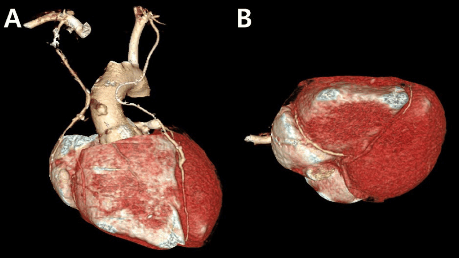
Patient 1 died of colon cancer with liver metastasis one year after the operation. Patient 2 had a history of preoperative mitral regurgitation and ventricular dysfunction, and prior RCA stent placement with subsequent in-stent restenosis happened. Postoperatively, patient 2 developed ventricular dysfunction (ejection fraction = 39%) and heart failure. Patient 3 is doing well 41.9 months after the operation, and computed tomography angiography performed one year after CABG demonstrated patent left and right ITA grafts (Fig. 1).
Conclusion
Coronary revascularization using ITA grafts is the gold standard treatment for KD-related coronary sequelae. Long-term patency, growth compatibility, and survival outcomes clearly favor arterial grafting over SVGs. While long-term survival exceeds 90% at 25–30 years, ventricular function at the time of intervention remains the strongest determinant of prognosis. Early ischemia detection, careful procedural selection, and lifelong surveillance are essential to optimizing outcomes due to the chronic and evolving nature of KD-related coronary sequelae. Ongoing refinement of surgical and postoperative management will further enhance outcomes in this unique patient population. As this population ages, collaboration between pediatric cardiology, cardiothoracic surgery, and adult congenital heart specialists will become increasingly critical.


