
Introduction
Kawasaki disease (KD) is acute systemic vasculitis predominantly affecting children, which can lead to significant cardiovascular complications. Among these complications, coronary artery abnormalities, including dilation and aneurysms, are the biggest concern due to ischemic heart disease [1]. In spite of advancements in the acute treatment of KD, some patients still progress to severe coronary complications, such as coronary artery giant aneurysm and even complete occlusion [2].
The management of coronary artery lesions in KD has traditionally involved both medical and interventional approaches, particularly when severe abnormalities are present [3]. However, the decision of optimal interventional timing is not easy even if the patient showed no ischemic symptoms and signs [4]. Additionally, the patient may exhibit the natural adaptive changes using collateral arteries that occur in coronary arteries over time.
This paper aims to share the feasibility and experiences of conservative monitoring in patients with KD who have developed total occlusion of the right coronary artery (RCA). By reviewing current literature and analyzing case studies, we would like to provide a framework for future management strategies in similar cases.
Methods
We reviewed patients who were diagnosed with KD and underwent a cardiac computed tomography (CT) between 2000 and 2022. In total, seventy-eight patients were identified in our institute, and among them, 7 patients showed RCA total occlusion. In our group, patients with simultaneously left coronary artery (LCA) occlusion or significant stenosis were excluded although patients with RCA total occlusion and LCA dilatation were enrolled.
Our institute recommends regular investigations with echocardiography once or twice a year, and annual cardiac CT or coronary angiography for patients who had a giant coronary artery dilatation after KD. The treadmill test or myocardial single-proton emission computed tomography (SPECT) are recommended if the patient complains about angina-like symptoms or has stenotic coronary lesions. For the long-term thromboprophylaxis, we recommend triple anti-thrombotic therapy with anticoagulation and dual antiplatelet agents in patients with large or giant coronary aneurysmal dilatation after KD by AHA guidelines [5].
Results
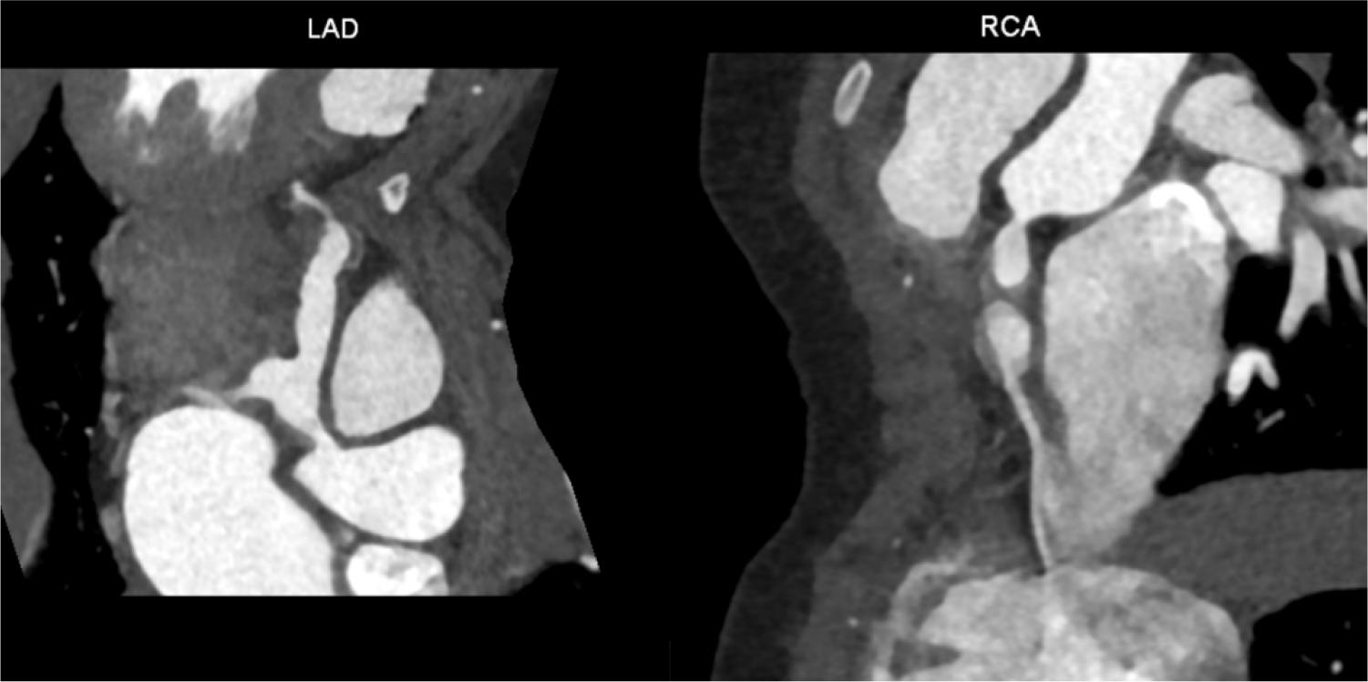
In total, seven patients were included in our study and their baseline characteristics were summarized in Table 1. Five patients were male and the age range of the total 7 patients at the time of KD diagnosis was from 1.3 to 8.5 years. Before diagnosis of RCA total occlusion, all patients had giant coronary artery dilatation by KD. The range of age at diagnosis of RCA total occlusion was from 1.8 to 11.0 years and median duration from KD to diagnosis of RCA total occlusion was 2.3 years (range from 0.5 to 5.7 years). The diagnosis of RCA total occlusion was made by cardiac CT (Fig. 1). In evaluation of LCA, 5 patients showed a giant aneurysmal dilatation in LCA and 2 patients showed intact LCA. Three patients showed mild chest pain, and 4 patients did not have any subjective symptoms.
After the diagnosis of RCA total occlusion, all patients underwent additional cardiac imaging or functional study such as coronary angiography, echocardiography, treadmill test, and myocardial perfusion SPECT. In evaluation for LCA, 5 patients showed a giant aneurysmal dilatation in LCA and 2 patients showed intact LCA. No patients showed ischemic findings in electrocardiogram and treadmill test although the latter was performed in only 5 patients. In echocardiography, no patients showed left ventricular wall motion limitation and only 1 patient showed a hyperechogenic change in left ventricular inferior wall. Before the diagnosis of RCA occlusion, all patients had taken 3 types of anti-thrombotic agents and the international normalized ratio (INR) of anti-coagulation was well maintained between 1.5 and 3.0. After RCA total occlusion diagnosis, we recommended the medical follow up and all patients have maintained 3 types of anti-thrombotic agents. In only two patients, subcutaneous low-molecular weight heparin was used transiently according to the opinion of attending physicians. Their median duration of follow up was 7.2 years (range 1.0–13.5 years) after RCA total occlusion and all patients showed New York Heart Association (NYHA) functional class I.
Discussion
Our results showed the long-term outcomes of conservative management in patients with RCA total occlusion following KD. Although the risk of ischemic heart disease is well analyzed in adults with an atherosclerotic coronary artery disease, insufficient data exists for that in pediatric patients with coronary artery complications after KD. Our findings suggest that a conservative approach, involving regular follow-up and medical therapy, may be a viable management option in selected patients who had RCA occlusion and did not have any ischemic signs. These results provide valuable insights into the long-term prognosis of patients with RCA occlusion and important considerations for clinical decision-making. In our institute, for pediatric patients with RCA total occlusion and no ischemic finding after KD, medical management is considered firstly.
One of the key findings of our study was that none of the patients exhibited significant ischemic changes on electrocardiography, treadmill tests, or myocardial perfusion SPECT. This aligns with previous research suggesting that the development of collateral circulation in KD may mitigate ischemic symptoms, even in cases of total occlusion [6]. Several studies have reported that spontaneous recanalization or progressive remodeling of coronary arteries can occur over time, particularly in pediatric patients, thereby reducing the immediate need for invasive revascularization strategies [7]. However, the exact mechanisms of these compensatory changes remain unclear. Some researchers hypothesize that endothelial progenitor cells and vascular endothelial growth factors play a role in collateral vessel formation [8]. Additionally, experimental models have suggested that arterial remodeling is influenced by hemodynamic forces, particularly in young patients with active angiogenic potential [9].
Despite the anatomical severity of total RCA occlusion, all patients in our cohort remained in NYHA functional class I. This suggests that adaptive coronary changes may play a crucial role in maintaining myocardial perfusion, allowing patients to maintain an acceptable quality of life without overt cardiac symptoms [8,10,11]. Some pediatric patients with severe coronary complications of KD can achieve long-term survival with medical management alone. However, subtle reductions in myocardial perfusion reserve cannot be completely ruled out. Recently, there are reports that advanced perfusion imaging techniques such as positron emission tomography (PET) or cardiac magnetic resonance imaging may provide more detailed insights [3,12]. Further longitudinal studies are needed to evaluate whether these patients remain asymptomatic into adulthood.
Another critical aspect of our study was the use of aggressive antithrombotic therapy, including aspirin, clopidogrel, and warfarin or low-molecular-weight heparin. Previous studies have demonstrated that patients with KD-related coronary artery abnormalities have heightened risk of thrombotic complications due to endothelial dysfunction and turbulent blood flow within aneurysmal segments [3,4,13]. The role of triple antithrombotic therapy remains debated in adult patients with atherosclerotic vascular lesions, but emerging evidence supports its use in high-risk patients with coronary abnormalities by KD to prevent adverse events [5,14]. However, concerns about the long-term safety of chronic anti-coagulation, particularly in pediatric populations, have been raised. Issues such as increased bleeding risk and medication adherence must be considered when making individualized treatment decisions.
Despite the promising findings of our study, several limitations should be acknowledged. The small sample size limits the generalizability of our results, and the retrospective nature of our analysis may introduce potential biases. Additionally, functional assessments such as coronary flow reserve measurements or stress imaging were not performed, potentially underestimating the burden of subclinical ischemia. Future studies should incorporate prospective designs, larger cohorts, and advanced imaging techniques to further elucidate the natural history and optimal management strategies for RCA total occlusion in KD. Research into the molecular mechanisms underlying vascular remodeling and potential biomarkers for ischemic risk stratification may help refine clinical management strategies [3,14].
Conclusion
Our study suggests that conservative management with regular follow-up and medical therapy can be an option for selected patients with RCA total occlusion following KD and without any ischemic findings. In this situation, the use of triple anti-thrombotic therapy was recommended for preventing adverse events. Further research is needed to establish standardized guidelines for the long-term management of these patients.