서론
후인두 염증(retropharyngeal inflammation, RPI)은 인두와 경추 사이에 위치하며 상부 종격동까지 확장되는 후인두 공간(retropharyngeal space)에 발생하는 염증을 가리킨다[1–3]. 후인두 주위의 림프절은 5세 이후에 퇴화하기 때문에, RPI는 3–4세 미만의 어린 소아에 호발한다[1]. RPI는 주로 상기도 감염 질환의 합병증 형태로 발생하지만, 심한 염증반응을 유발하는 비감염 원인에 의해서도 발생할 수 있다[2,3]. RPI는 심한 정도에 따라, 부종(edema), 연조직염(cellulitis) 또는 플레그몬(phlegmon), 화농(suppuration) 또는 농양 순으로 구분한다[4,5]. 가장 심한 형태인 후인두 농양(retropharyngeal abscess, RPA)은 흔하지는 않지만 기도 폐쇄나 종격동염(mediastinitis)을 초래할 수 있으므로, 조기에 진단하여 적극적으로 치료하는 것이 중요하다[6]. 하지만 대부분의 RPA 소아환자는 발열, 보챔, 식욕부진 등 비특이적 증상을 나타내어 조기 인식이 어려울 수 있다.
가와사키병(Kawasaki disease, KD)은 5세 미만 소아에 호발하는 원인 미상의 전신 혈관염이다[7,8]. KD는 발열과 진단기준에 포함되는 5가지 주요 임상증상(결막염, 구인두의 염증, 발진, 손발의 변화 및 경부 림프절종대)을 특징으로 한다. 드물지만, 일부 환자는 KD의 주요 임상증상 없이 RPI를 주 증상으로 나타내기도 한다[9,10]. RPI가 동반된 KD (KD with RPI, RPI-KD)는 세균 RPA로 오인될 수 있으며, 이로 인해 KD의 진단과 치료가 지연되어 관상동맥 이상(coronary artery abnormalities, CAAs)의 위험도가 증가할 수 있다[10,11]. 본 연구에서 저자들은 문헌고찰을 통해 RPI-KD의 임상양상, 영상의학 소견, 치료 및 임상경과를 조사하고 RPI-KD와 세균 RPA를 구분하는 데 도움이 되는 진단적 단서를 확인하려고 한다.
본론
PubMed에서 핵심용어(keywords)를 이용하여 RPI-KD에 대한 문헌을 검색하였다(Kawasaki disease or mucocutaneous lymph node syndrome) AND (retropharyngeal abscess or deep neck infection). 전체 36편의 논문이 검색되었다. 이 중 대상 환자 4명 이하의 증례 보고 24편, 종설 5편, 소아다기관염증증후군(multisystem inflammatory syndrome in children, MIS-C)에 대한 사설 1편을 제외하고, 최종적으로 원저 6편이 본 문헌고찰 연구를 위해 선택되었다[11–16]. Inagaki et al.[16]의 연구를 제외한 나머지 연구에서 모든 RPI-KD 환자들은 경부 computed tomography (CT) 검사를 시행받았다. Chen et al.[11]의 연구는 3개 환자군, 즉 RPI-KD군, RPI가 동반되지 않은 KD군 및 세균 RPA군의 특징을 비교하였고 그 외 연구들은 2개 환자군, 즉 RPI-KD군과 세균 RPA군 또는 RPI-KD군과 KD군의 특징을 비교하였다. Table 1에는 본 연구에 포함된 원저 6편의 추가적인 정보를 요약하였다.
| Authors, reported year | Country | Methods1) | Study population | ||
|---|---|---|---|---|---|
| RPI-KD (n) | KD (n) | Bacterial RPA (n) | |||
| Chen et al. [11], 2022 | China | Single center | 10 | 20 | 16 |
| Lim et al. [12], 2020 | Korea | Single center | 11 | – | 36 |
| Liu et al. [13], 2024 | China | Single center | 22 | – | 34 |
| Nomura et al. [14], 2014 | Japan | Single center | 21 | – | 18 |
| Tona et al. [15], 2014 | Japan | Single center | 10 | 267 | – |
| Inagaki et al. [16], 2019 | United States | Insurance data | 130 | 20,657 | – |
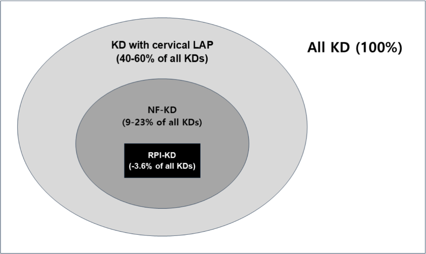
Fig. 1에는 (i) 경부 림프절종대 동반 KD, (ii) NF-KD (node-first presentation of Kawasaki disease) 및 (iii) RPI-KD의 상대적인 빈도를 나타냈다. 경부 림프절종대는 진단기준에 포함되는 주요 임상증상으로, KD 환자의 약 40%–60%에서 관찰된다[17,18]. NF-KD는 질병의 초기에 다른 KD의 주요 임상증상 없이 경부 림프절종대를 주 증상 또는 유일한 증상으로 나타내는 KD를 일컫는다[4]. NF-KD는 KD 환자의 9%–23%에서 보고되었다[9,19]. RPI-KD가 세균 RPA로 오인될 수 있는 것처럼, NF-KD는 세균 경부 림프절염(bacterial cervical lymphadenitis, BCL)과 혼돈될 수 있다[19,20]. RPI-KD는 KD 환자의 0.6%–3.6%에서 발생하는 것으로 보고되었다[15,16].

Table 2에는 RPI-KD군, KD군(즉, RPI 동반 없는 KD), 세균 RPA군의 임상적 특징을 나타냈다. 남아의 비율은 3군 모두에서 높았으며, 평균 연령은 RPI-KD군과 세균 RPA군에서 유사하였고 KD군에서는 통계적으로 유의하게 낮았다. 발열 기간은 대부분의 연구에서 유사했으나, Lim et al.[12]의 연구에서는 세균 RPA군보다 RPI-KD군에서 길었다. 이를 근거로, Lim et al.[12]은 항생제 치료 시작 3일 후에도 발열이 지속되는 RPA 환자에서 RPI-KD 가능성을 고려해야 한다고 강조하였다. 발열과 보챔과 같은 비특이적 증상과 함께, 경부 통증, 연하곤란(dysphagia), 경부 운동 제한(limited motion of neck), 경부 강직(neck stiffness), 협착음(stridor), 호흡곤란 등의 추가 증상이 3군 모두에서 관찰되었다. 경부 통증과 연하곤란은 RPI-KD군이나 KD군보다 세균 RPA군에서 흔했다[13,14].
| Variables | RPI-KD | KD | Bacterial RPA | References | |||
|---|---|---|---|---|---|---|---|
| Male sex (%) | 50.0 | (n = 10) | 60.0 | (n = 20) | 56.3 | (n = 16) | [11] |
| 81.8 | (n = 11) | – | 58.3 | (n = 36) | [12] | ||
| 52.9 | (n = 22) | – | 54.5 | (n = 34) | [13] | ||
| 66.7 | (n = 21) | – | 55.5 | (n = 18) | [14] | ||
| 60.0 | (n = 10) | 61.8 | (n = 267) | – | [15] | ||
| 67.9 | (n = 130) | 59.3 | (n = 20,657) | – | [16] | ||
| Age (years) | 5.5 | (n = 10) | 2.61) | (n = 20) | 4.7 | (n = 16) | [11] |
| 4.0 | (n = 11) | – | 4.0 | (n = 36) | [12] | ||
| 5.6 | (n = 22) | – | 5.7 | (n = 34) | [13] | ||
| 5.0 | (n = 21) | – | 4.5 | (n = 18) | [14] | ||
| 4.5 | (n = 10) | 1.01) | (n = 267) | – | [15] | ||
| 5.0 | (n = 130) | 2.01) | (n = 20,657) | – | [16] | ||
| Duration of fever (days) | 7.0 | (n = 10) | 7.1 | (n = 20) | 5.4 | (n = 16) | [11] |
| 6.01) | (n = 11) | – | 5.0 | (n = 36) | [12] | ||
| 10.1 | (n = 22) | – | 8.7 | (n = 34) | [13] | ||
| 5.0 | (n = 21) | – | 5.0 | (n = 18) | [14] | ||
| 4.5 | (n = 10) | 4.0 | (n = 267) | – | [15] | ||
| Neck pain (%) | 100.0 | (n = 10) | 25.01) | (n = 20) | 100.0 | (n = 16) | [11] |
| 36.4 | (n = 11) | – | 44.4 | (n = 36) | [12] | ||
| 81.8 | (n = 22) | – | 79.4 | (n = 34) | [13] | ||
| 57.12) | (n = 21) | – | 94.4 | (n = 18) | [14] | ||
| Dysphagia (%) | 60.0 | (n = 10) | 0.01) | (n = 20) | 68.7 | (n = 16) | [11] |
| 9.1 | (n = 11) | – | 25.0 | (n = 36) | [12] | ||
| 0.02) | (n = 22) | – | 17.6 | (n = 34) | [13] | ||
| 23.82) | (n = 21) | – | 61.1 | (n = 18) | [14] | ||
| Limited motion of neck (%) | 100.0 | (n = 10) | 25.01) | (n = 20) | 93.7 | (n = 16) | [11] |
| 45.5 | (n = 11) | – | 47.2 | (n = 36) | [12] | ||
| 63.6 | (n = 22) | – | 64.7 | (n = 34) | [13] | ||
| 52.4 | (n = 21) | – | 83.3 | (n = 18) | [14] | ||
Table 3에는 RPI-KD군, KD군, 세균 RPA군의 검사실 결과를 나타냈다. 공통적인 혈액검사 항목에는 혈색소, 백혈구, 혈소판, 적혈구 침강 속도(erythrocyte sedimentation rate, ESR), C반응 단백(C-reactive protein, CRP), aspartate aminotransferase (AST), alanine aminotransferase (ALT)이 포함되었다. CRP 값은 3군 중 RPI-KD군에서 가장 높았고[11], AST와 ALT 값은 세균 PRA군보다 RPI-KD군에서 높았다[13,15]. 혈색소, 혈소판, ESR 값은 3군에서 유의한 차이가 없었고, 백혈구 값은 연구[11,15]에 따라 상반된 결과를 보였다. Chen et al.[11]의 연구에서 추가로 프로칼시토닌(procalcitonin, PCT), N말단 뇌나트륨이뇨펩티드(N-terminal pro-brain natriuretic peptide, NT-proBNP), 트로포닌(troponin) 검사를 시행하였고, 이중 트로포닌 값은 세균 PRA군보다 RPI-KD군에서 유의하게 높았다. 즉, 트로포닌의 상승은 세균 RPA군에서 관찰되지 않는 RPI-KD군 특이적 소견이었다. 세균 RPA군에서 시행되지 않은 혈액검사 항목(NT-proBNP와 페리틴)에 대한 통계적 유의성은 확인할 수 없었다.
| Variables | RPI-KD | KD | Bacterial RPA | References | |||
|---|---|---|---|---|---|---|---|
| Hb (g/dL) | 11.6 | (n = 10) | 11.1 | (n = 20) | 11.6 | (n = 16) | [11] |
| 11.8 | (n = 11) | – | 11.8 | (n = 36) | [12] | ||
| 11.3 | (n = 22) | – | 11.8 | (n = 34) | [13] | ||
| WBC (× 109/L) | 16.3 | (n = 10) | 14.9 | (n = 20) | 20.82) | (n = 16) | [11] |
| 15.7 | (n = 11) | – | 16.9 | (n = 36) | [12] | ||
| 18.0 | (n = 22) | – | 19.9 | (n = 34) | [13] | ||
| 20.9 | (n = 21) | – | 22.3 | (n = 18) | [14] | ||
| 21.01) | (n = 10) | 13.3 | (n = 267) | – | [15] | ||
| Platelet (× 109/L) | 352 | (n = 10) | 327 | (n = 20) | 412 | (n = 16) | [11] |
| 308 | (n = 11) | – | 342 | (n = 36) | [12] | ||
| 333 | (n = 22) | – | 394 | (n = 34) | [13] | ||
| ESR (mm/hr) | 64 | (n = 10) | 55 | (n = 20) | 66 | (n = 16) | [11] |
| 67 | (n = 11) | – | 60 | (n = 36) | [12] | ||
| 85 | (n = 22) | – | 67 | (n = 34) | [13] | ||
| CRP (mg/dL) | 9.41) | (n = 10) | 5.2 | (n = 20) | 5.8 | (n = 16) | [11] |
| 9.7 | (n = 11) | – | 9.7 | (n = 36) | [12] | ||
| 10.1 | (n = 22) | – | 7.2 | (n = 34) | [13] | ||
| 11.7 | (n = 21) | – | 9.9 | (n = 18) | [14] | ||
| 11.12) | (n = 10) | 5.6 | (n = 267) | – | [15] | ||
| AST (U/L) | 662) | (n = 10) | 70 | (n = 20) | 25 | (n = 16) | [11] |
| 26 | (n = 11) | – | 26 | (n = 36) | [12] | ||
| 522) | (n = 22) | – | 28 | (n = 34) | [13] | ||
| 27 | (n = 21) | – | 24 | (n = 18) | [14] | ||
| ALT (U/L) | 911) | (n = 10) | 104 | (n = 20) | 14 | (n = 16) | [11] |
| 13 | (n = 11) | – | 14 | (n = 36) | [12] | ||
| 342) | (n = 22) | – | 16 | (n = 34) | [13] | ||
| PCT (ng/mL) | 1.2 | (n = 10) | 2.8 | (n = 20) | 0.5 | (n = 16) | [11] |
| 0.6 | (n = 22) | – | 0.4 | (n = 34) | [13] | ||
| NT-proBNP (ng/L) | 248 | (n = 10) | 175 | (n = 20) | – | [11] | |
| Ferritin (ng/mL) | 490 | (n = 10) | 260 | (n = 20) | – | [11] | |
| Troponin (ng/mL) | 1.9 | (n = 10) | 4.3 | (n = 20) | 0.52) | (n = 16) | [11] |
RPI-KD: Kawasaki disease with retropharyngeal inflammation; KD: Kawasaki disease; RPA: retropharyngeal abscess; Hb: hemoglobulin; WBC: white blood cell counts; ESR: erythrocyte sedimentation rate; CRP: C-reactive protein; AST: aspartate aminotransferase; ALT: alanine aminotransferase; PCT: procalcitonin; NT-proBNP: N-terminal pro-brain natriuretic peptide.
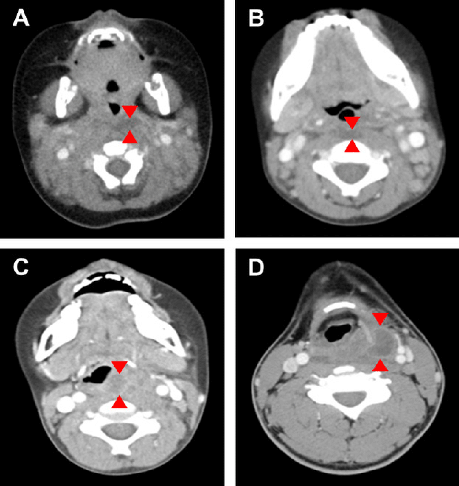
Table 4에는 RPI-KD군과 세균 RPA군의 경부 CT 검사 결과를 비교하였다. 연조직염 또는 플레그몬의 빈도는 RPI-KD군에서 높았고, 화농 또는 농양의 빈도는 세균 RPA군에서 높았다[12,13,15]. 하지만, 화농 또는 농양 병변은 세균 RPA군에만 나타난 것이 아니고, RPI-KD군의 45.5%에서도 관찰되었다[13]. 이는 CT 검사에서 화농 또는 농양 병변이 확인되었더라도 RPI-KD를 완전히 배제시킬 수 없다는 것을 의미한다. RPI-KD군과 세균 RPA군 간의 가장 두드러진 차이점은 고리 또는 테두리 조영증강(ring or rim enhancement)의 유무였다. 고리 조영증강은 세균 RPA군(88.9%–100.0%)에서는 흔하게 확인되었으나, RPI-KD군(0.0%)에서는 관찰되지 않았다[11,14,15]. 즉, 고리 조영증강은 RPI-KD군에서 관찰되지 않는 세균 RPA군 특이적 소견이었다. 종괴 효과(mass effect)도 RPI-KD군(4.7%)보다 세균 RPA군(61.1%)에서 빈번하게 관찰되었다[14]. Fig. 2에는 RPI-KD 환자에서 확인된 연조직염 또는 플레그몬의 CT 영상(A, B)과 세균 RPA 환자에서 확인된 고리 조영증강이 동반된 화농 또는 농양의 CT 영상(C, D)을 나타냈다.
| Variables | RPI-KD | Bacterial RPA | References | ||
|---|---|---|---|---|---|
| Cellulitis or phlegmon (%) | 100.01) | (n = 11) | 80.6 | (n = 36) | [12] |
| 81.81) | (n = 22) | 44.1 | (n = 34) | [13] | |
| 100.0 | (n = 10) | – | [15] | ||
| Suppuration or abscess (%) | 0.01) | (n = 11) | 44.4 | (n = 36) | [12] |
| 45.51) | (n = 22) | 85.3 | (n = 34) | [13] | |
| Ring enhancement (%) | 0.01) | (n = 10) | 100.0 | (n = 16) | [11] |
| 0.01) | (n = 21) | 88.9 | (n = 18) | [14] | |
| 0.0 | (n = 10) | – | [15] | ||
| Mass effect (%) | 4.71) | (n = 21) | 61.1 | (n = 18) | [14] |

Table 5에는 RPI-KD군, KD군, 세균 RPA군의 치료와 임상경과를 요약하였다. RPI-KD군과 세균 RPA군의 모든 환자는 항생제를 치료를 받았고, 절반 이상의 환자는 두 종류 이상의 항생제 치료를 받았다[12,13]. 세균 RPA군의 약 20% 환자는 수술적 치료가 필요했고, RPI-KD군 중 일부(9.1%) 환자도 세균 RPA로 오인되어 수술적 치료를 받았다[12,13]. 치료 방법 측면에서, RPI-KD군과 세균 RPA군 간의 유의한 차이는 없었다.
입원기간은 3군 중 RPI-KD군에서 가장 길었고, 의료비 역시 KD군보다 RPI-KD군에서 더 많이 지출되었다[11,16]. 이는 RPI-KD 환자들이 세균 RPA로 오인되어 KD의 진단은 지연되고 비필수적인 검사와 치료를 받았기 때문이라고 여겨진다. 일반적으로 RPI-KD의 전신염증이 KD보다 심하다고 알려져 있으나, 일부 연구[15,16]에서는 CAAs가 KD군보다 RPI-KD군에서 낮은 빈도로 확인되었다.
| Variables | RPI-KD | KD | Bacterial RPA | References | |||
|---|---|---|---|---|---|---|---|
| Abx, single (%) | 100.0 | (n = 11) | – | 100.0 | (n = 36) | [12] | |
| Abx, multiple (%) | 54.5 | (n = 22) | – | 55.9 | (n = 34) | [13] | |
| Surgery (%) | 0.0 | (n = 11) | – | 16.7 | (n = 36) | [12] | |
| 9.1 | (n = 22) | – | 14.7 | (n = 34) | [13] | ||
| Length of stay (days) | 11.11) | (n = 10) | 6.7 | (n = 20) | 8.6 | (n = 16) | [11] |
| 7.01) | (n = 130) | 3.0 | (n = 20,657) | – | [16] | ||
| Medical cost ($) | 19,7731) | (n = 130) | 8,645 | (n = 20,657) | – | [16] | |
| Coronary artery | 20.0 | (n = 10) | 5.0 | (n = 20) | – | [11] | |
| complications (%) | 0.0 | (n = 10) | 3.7 | (n = 267) | – | [15] | |
| 1.1 | (n = 130) | 3.8 | (n = 20,657) | – | [16] | ||
NF-KD는, 염증의 심한 정도나 영상의학 소견과 무관하게, 경부 림프절종대를 주 증상으로 나타내는 KD로 정의된다는 점에서 근본적으로 RPI-KD와는 다르다. 하지만 RPI-KD와 유사하게, NF-KD는 일반적인 KD보다는 심한 전신염증을 나타내며 경부 CT 검사에서 이상을 보이는 경우가 적지 않다[21–24]. 최근 발표된 NF-KD와 BCL의 비교 연구[9,21]에서, NF-KD에서 BCL보다 평균 연령이 높았고 발열 기간도 길었다. 또한 BCL에 비해, NF-KD에서 빈혈, 백혈구증가증, ESR과 CRP 증가, AST와 ALT 증가, 저나트륨혈증, NT-proBNP 증가와 같은 검사실 이상 소견이 흔하였다.
RPI-KD 환자와 다르게, 모든 NF-KD 환자에서 영상검사가 시행되지는 않지만, 후인두 공간의 감염이 의심되거나 진단이 모호한 경우에는 영상검사를 시행한다. 예를 들어, Kanegaye et al. [4]의 연구에서 NF-KD 환자의 21%(12/57)는 경부 CT 또는 초음파 검사를 시행받았다. You [9]는 문헌고찰을 통해 NF-KD 환자와 BCL 환자의 영상 소견을 비교하였다. 경부 CT 검사에서 군집 림프절(clustered lymph nodes), 부종과 연조직염은 NF-KD 환자에서 흔하였고, 우세 종괴(dominant mass)와 화농 또는 농양은 BCL 환자에서 흔하였다[22,23]. 이와 유사하게, 초음파 검사에서도 군집 림프절과 이질적 에코음영(echogenicity)은 NF-KD 환자에서 흔하였고, 우세 종괴, 동질적 에코음영, 불규칙 경계 및 화농 또는 농양은 BCL 환자에서 흔하였다[23,24].
인구학적 특징 비교에서 RPI-KD군과 세균 RPA군 간의 유의한 차이는 없었다. 임상증상 중 경부 통증과 연하곤란은 세균 RPA군에서 좀 더 흔하게 관찰되었다. 검사실 소견에서 RPI-KD군은 세균 RPA군보다 높은 CRP, AST, ALT, 트로포닌 값을 보였으며, 트로포닌 상승은 RPI-KD군에서만 관찰되었다. 경부 CT 검사 비교에서 연조직염 또는 플레그몬은 RPI-KD군에 흔하였고, 화농 또는 농양은 세균 RPA군에 흔했다. 고리 조영증강은 세균 RPA군에는 흔했지만, RPI-KD군에서는 확인되지 않았다. RPI-KD군의 모든 환자는 경험적 항생제 치료를 받았고, 일부 환자는 수술적 치료를 받았다. KD군과 비교하여, RPI-KD군에서 입원기간이 길었고 더 많은 의료비가 지출되었다.
결론
실제 임상에서 RPI-KD 환자가 세균 RPA로 오인되어 비필수적인 검사와 치료를 받을 수 있다. CRP, AST 및 ALT의 증가는 RPI-KD에서 상대적으로 흔하지만, 세균 RPA가 악화되었을 때도 보일 수 있다. 마찬가지로, CT 검사에서 화농 또는 농양은 세균 RPA에서 흔하지만, 심한 형태의 RPI-KD에서도 관찰될 수 있다. 본 연구에서 조사한 항목 중 혈액검사의 트로포닌 증가는 RPI-KD에 특이적이었으며, CT 검사의 고리 조영증강은 세균 RPA에 특이적이었다. 그 밖의 항목들은 RPI-KD 또는 세균 RPA에서만 관찰되는 특징은 아니었다. 따라서 RPI-KD를 간과하지 않기 위해서는 적절한 항생제 치료에도 불구하고 임상적 호전이 없는 세균 RPA 환자에서, RPI-KD의 가능성을 고려하는 것이 중요하다.
